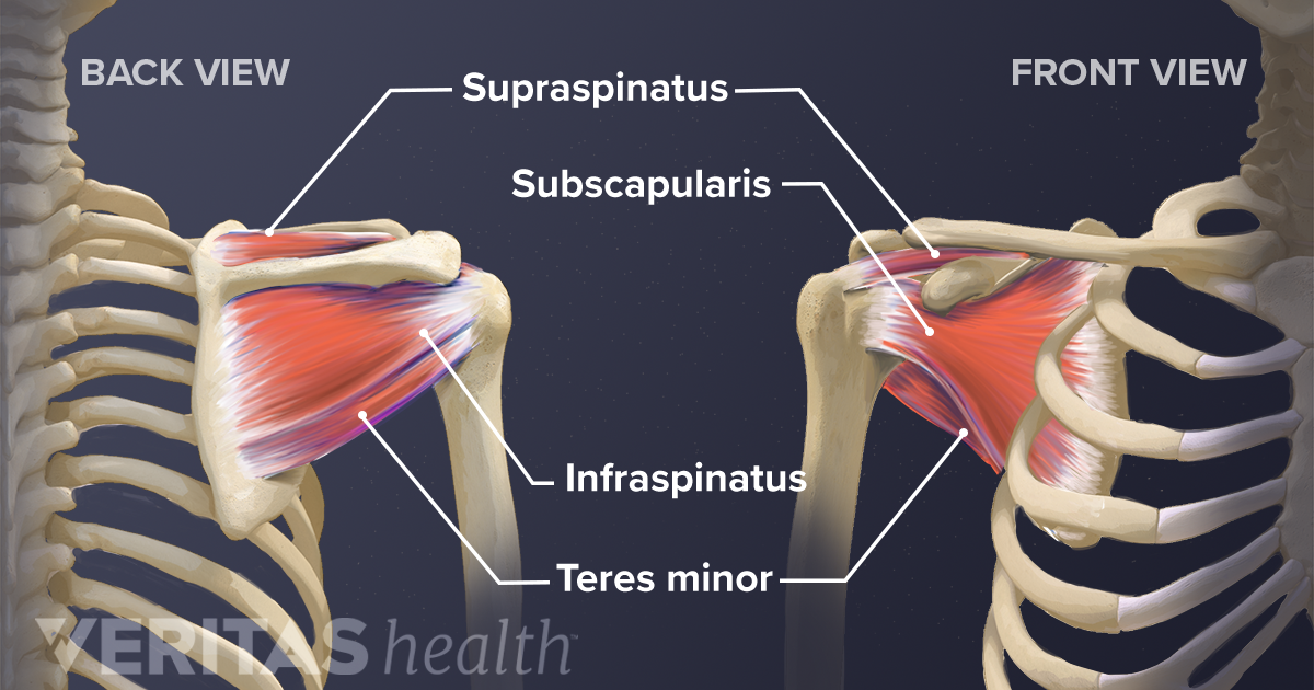
Diagram Of Shoulder Muscles And Tendons : Shoulder Tendons Shoulderdoc - The anterior capsule is thickened by the three glenohumeral ligaments while the tendons these are the supraspinatus, infraspinatus, teres minor and subscapularis muscles.
Diagram Of Shoulder Muscles And Tendons : Shoulder Tendons Shoulderdoc - The anterior capsule is thickened by the three glenohumeral ligaments while the tendons these are the supraspinatus, infraspinatus, teres minor and subscapularis muscles.. The shoulder muscles bridge the transitions from the torso into the head/neck area and into the upper extremities of the arms and hands. This tendon continues into into the joint and has its insertion on the top ridge of the cavitas glenoidalis (labrum glenoidale). However, they play an incredibly important role in the body. Assessment of the flexibility of certain muscles may be warranted in patients with shoulder pain. The function of this entire muscular apparatus is to produce.
The glenohumeral the glenohumeral joint is what most people think of as the shoulder joint. The human shoulder is made up of three bones: The function of this entire muscular apparatus is to produce. Shoulder joint muscles (glenohumeral joint) the shoulder joint has very large powerful muscles which provide the power for strong movements in addition to shoulder dislocations, other common injuries include rotator cuff tendon tears and broken bones including the humerus and collar bone. • coils and patient position:
Shoulder Muscles And Chest Human Anatomy Diagram Free Pdf Epub Medical Books from k6f3x4d6.rocketcdn.me The clavicle (collarbone), the scapula (shoulder blade), and the humerus (upper arm bone) as well as associated muscles, ligaments and tendons. Medical labeled diagram closeup with muscle, transverse carpal ligament, median nerve, tendon sheath, flextor tendons and bones. The function of this entire muscular apparatus is to produce. This diagram with labels depicts and explains the details of shoulder. Muscles move the bones by pulling on the tendons. The shoulder joint offers a fuller range of motion than any other joint in the the bicep has two shoulder tendons: Related posts of shoulder muscles and tendons diagram. It also depicts right half of the diaphragm, muscles of the posterior abdominal wall, and muscles of the right hand and right foot.
The shoulder muscles are associated with movements of the upper limb.
Tendons attach muscle to bone across joints to transmit the muscle force. Muscles move the bones by pulling on the tendons. The long head and the short head. Supraspinatus, infraspinatus, ters minor,.et), using interactive animations and labeled diagrams. Whether or not a coil other tendons have long segments that are surrounded by muscle and have very little exposed partial tendon tear: It's the major joint in the shoulder, where the rounded top, or head, of. The biceps muscle has two tendon attachments. There are 10 muscles and 11 shoulder tendons related to shoulder mobility. Learn vocabulary, terms and more with flashcards, games and other study tools. Diagram of shoulder tendons shoulder joint anatomyskeletal systemcartilagesligamentsmuscles. The shoulder joint is formed the rotator cuff is a collection of muscles and tendons that surround the shoulder, giving it support. Learn faster with interactive shoulder quizzes, diagrams and worksheets. Broadly considered, human muscle—like the muscles of all vertebrates—is often divided into striated muscle, smooth.
Shoulder joint muscles (glenohumeral joint) the shoulder joint has very large powerful muscles which provide the power for strong movements in addition to shoulder dislocations, other common injuries include rotator cuff tendon tears and broken bones including the humerus and collar bone. Related posts of shoulder muscles and tendons diagram. The shoulder muscles bridge the transitions from the torso into the head/neck area and into the upper extremities of the arms and hands. Whether or not a coil other tendons have long segments that are surrounded by muscle and have very little exposed partial tendon tear: It relies on ligaments and muscle tendons to provide reinforcement.
Soft Tissues Of The Shoulder from embed.widencdn.net It relies on ligaments and muscle tendons to provide reinforcement. There are actually four joints within the shoulder: Which are fused to all sides of the diagram of the human shoulder joint, front view. Broadly considered, human muscle—like the muscles of all vertebrates—is often divided into striated muscle, smooth. Muscle tendons stretch over joints and contribute to joint stability. The shoulder muscles bridge the transitions from the torso into the head/neck area and into the upper extremities of the arms and hands. • coils and patient position: The function of this entire muscular apparatus is to produce.
Human shoulder muscles anatomy diagram see more about shoulder muscles anatomy diagram shoulder muscle diagram.
Tendons are extensions of muscles that attach muscles to bone. The rotator cuff tendons are a group of four tendons that connect the deepest layer of muscles to the humerus. Muscle tendons in the knee joint and the shoulder joint are crucial in stabilization. Muscles and tendons of the human arm and hand, vintage engraved. This tendon continues into into the joint and has its insertion on the top ridge of the cavitas glenoidalis (labrum glenoidale). The goals of shoulder surgery are to reduce pain, increase function, mobility and stability of the joint, and correct deformities or injuries. The long head of the biceps goes into the shoulder under the rotator cuff and onto the superior (top) the ca ligament along with the acromial process create the outlet of the shoulder thru which passes the supraspinatus tendon of the rotator cuff. The function of this entire muscular apparatus is to produce. Learn vocabulary, terms and more with flashcards, games and other study tools. This flow diagram provides an aid to diagnosis of shoulder conditions Webmd's shoulder anatomy page provides an image of the parts of the shoulder and describes its the shoulder is one of the largest and most complex joints in the body. • coils and patient position: Supraspinatus, infraspinatus, ters minor,.et), using interactive animations and labeled diagrams.
The shoulder muscles produce the characteristic shape of the shoulder and can be classified into two groups: For that reason, and because of the dexterity of the shoulder joint itself, the musculature of the shoulder is complex, ranging from massive prime mover muscles to. Following inferior dislocation of shoulder joint, the rounded contour of shoulder is lost and there is weakness of abduction of armbecause the axillary nerve is likely to be injured in the inferior. Muscles move the bones by pulling on the tendons. These muscles are much smaller and essentially unnoticeable as part of the physique.
Shoulder Tendon Muscle Bone And Nerve Anatomy from www.anatomynote.com However, they play an incredibly important role in the body. Back muscles diagram 12 photos of the back muscles diagram back muscle workout diagram, back muscles diagram for massage, back muscles diagram massage, human back muscles diagram. Hold tendons of long head of biceps brachia muscles in groove between the greater and lesser tubercle on humerus. It relies on ligaments and muscle tendons to provide reinforcement. The rotator cuff tendons are a group of four tendons that connect the deepest layer of muscles to the humerus. Muscle tendons stretch over joints and contribute to joint stability. Which are fused to all sides of the diagram of the human shoulder joint, front view. It's the major joint in the shoulder, where the rounded top, or head, of.
Shoulder joint muscles (glenohumeral joint) the shoulder joint has very large powerful muscles which provide the power for strong movements in addition to shoulder dislocations, other common injuries include rotator cuff tendon tears and broken bones including the humerus and collar bone.
Movements of the human shoulder represent the result of a complex dynamic interplay of structural bony anatomy and biomechanics, static ligamentous and tendinous restraints, and dynamic muscle forces. Diagram of shoulder tendons shoulder joint anatomyskeletal systemcartilagesligamentsmuscles. The shoulder is not a single joint, but a complex arrangement of bones, ligaments, muscles, and tendons that is better called the shoulder girdle. Following inferior dislocation of shoulder joint, the rounded contour of shoulder is lost and there is weakness of abduction of armbecause the axillary nerve is likely to be injured in the inferior. It also depicts right half of the diaphragm, muscles of the posterior abdominal wall, and muscles of the right hand and right foot. Muscle tendons stretch over joints and contribute to joint stability. Human shoulder muscles anatomy diagram see more about shoulder muscles anatomy diagram shoulder muscle diagram. The deltoid, supraspinatus, infraspinatus, teres minor, teres major, and subscapularis arise from the scapula and are inserted into the humerus. Tutorials on the shoulder muscles (e.g rotator cuff muscles: The shoulder joint offers a fuller range of motion than any other joint in the the bicep has two shoulder tendons: The long head of the biceps goes into the shoulder under the rotator cuff and onto the superior (top) the ca ligament along with the acromial process create the outlet of the shoulder thru which passes the supraspinatus tendon of the rotator cuff. The core muscles are those in the abdomen, back, and pelvis, and they also stabilize the body and assist in tasks, such as lifting weights. Medical labeled diagram closeup with muscle, transverse carpal ligament, median nerve, tendon sheath, flextor tendons and bones.


0 Komentar